
TIVITA®
Breakthrough technology for fast, accurate wound
diagnostics and wound healing evaluation.
The technology.
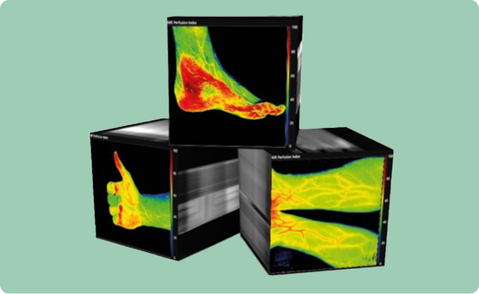
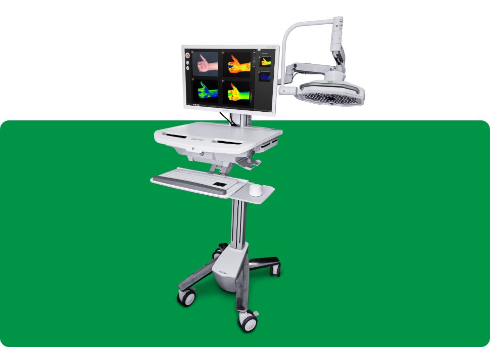
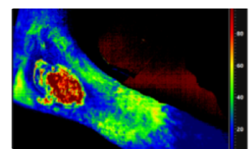
TIVITA® works by means of 100 different light channels between 500 and 1,000 nm. In 6.4 seconds TIVITA® allows the measurement of superficial oxygen saturation (StO2 [%], 1 mm in the skin), the deeper perfusion (NIR, 3 to 6 mm into the skin), the Tissue Hemoglobin index (THI) and the Tissue Water Index (TWI). This non-contact diagnostic tool not only allows to monitor the wound bed, but also the wound bed surroundings that often contain the reasons of bad or slower wound healing of complex and other wounds. The technology was recently developed based on imaging, spectroscopy, and tissue oximetry know-how.
TIVITA® exists in TIVITA® Tissue and TIVITA® Wound versions and is available both with cable and battery power supply. For every wound measurement done, a full series of values is available for all measured parameters.
TIVITA® for vascular surgeons
TIVITA® is a quick scanning tool for vascular or arterial occlusion assessment in the upper layers of the skin. In case problems are detected with TIVITA®, a more in-depth analysis with duplex Doppler can be provided. TIVITA® also helps in the quick assessment of revascularizations, the prevention or reduction of amputations by means of better, faster, and more complete assessments of the problem zones in DFU, VLU or pressure sores.
TIVITA® for plastic surgeons
Judging if donor sites or flaps will be successful is not an easy assessment to be done based on the limitations of the human eye. TIVITA® allows to see up to 6 mm deep into the skin and can easily be used to monitor the wound healing efficacy and efficiency of flaps and donor sites, to prevent them from getting lost due to oedema or perfusion problems that are invisible for the human eye or even for standard existing clinical assessment.
How it works.
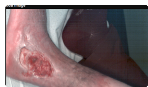
1.
RGB image (color image)
2.
St)2 Oxygenation image
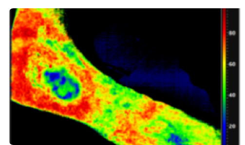
3.
NIR Perfusion image
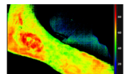
4.
TWI image
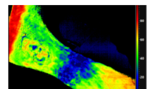
5.
THI image
Product brochures and instructions.
Request our TIVITA® product brochures and instructions for use here.
Discover our complementary product range.
Protex Healthcare offers a range of advanced, innovative products for the healing and closing of complex wounds.
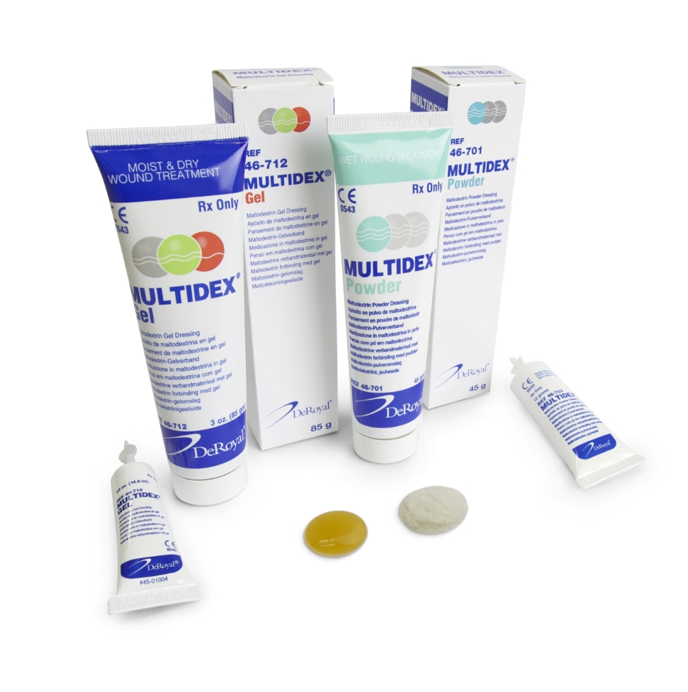
Multidex®
A wound dressing in gel or powder form, based on Maltodextrin and Vitamin C, for the treatment of complex wounds.
Learn more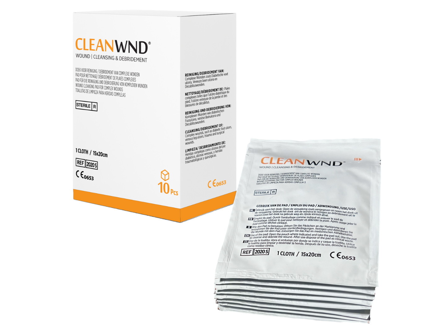
CleanWND®
Ready to use cloths for easy debridement and cleaning of the wound and the wound environment.
Learn more
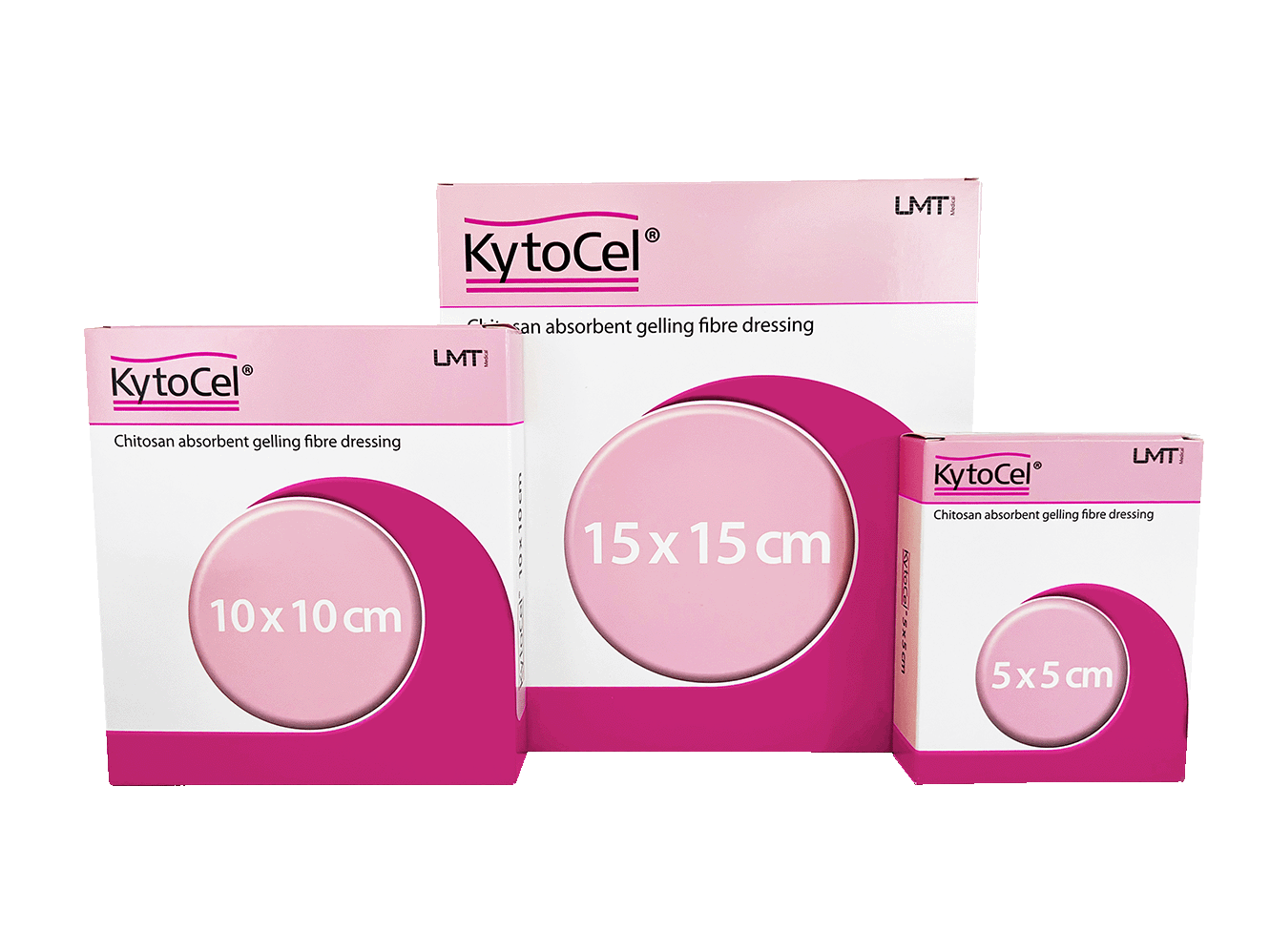
KytoCel®
KytoCel® is a highly absorbent, antimicrobial wound dressing with haemostatic, analgesic and scar-improving properties.
Learn more